DOI:
https://doi.org/10.14483/23448350.18500Published:
05/01/2022Issue:
Vol. 44 No. 2 (2022): May-August 2022Section:
Science and EngineeringDiagnosis of Gastrointestinal Parasites in Bovines of the Department of Boyacá, Colombia
Diagnóstico de parásitos gastrointestinales en bovinos del departamento de Boyacá, Colombia
Keywords:
Parasitic Diseases, Parasitology, helminths, nematodes, prevalence (en).Keywords:
Enfermedades de los bovinos, Parasitología, helmintos, nematodos, prevalencia (es).Downloads
References
Aram, A. (2020). Coprological study of gastrointestinal parasites in dairy cattle in sulaymaniyah province, kurdistan region, Iraq. Applied Ecology and Environmental Research, 18(5), 7279-2782. https://doi.org/10.15666/aeer/1805_72797287 DOI: https://doi.org/10.15666/aeer/1805_72797287
Ayele, A., Abay, M., Birhan, M., Yayeh, M., Erara, M., Gessese, T., Mohammed, A., Demoze, G. (2020). Prevalence of bovine gastrointestinal parasitic infection in and around Kombolcha Town. Online Journal of Animal and Feed Research, 10(2), 59-65. https://doi.org/10.36380/scil.2020.ojafr DOI: https://doi.org/10.36380/scil.2020.ojafr8
Bennema, S. C., Vercruysse, J., Morgan, E., Stafford, K., Höglund, J., Demeler, J., von Samson-Himmelstjerna, G., Charlier, J. (2010). Epidemiology and risk factors for exposure to gastrointestinal nematodes in dairy herds in northwestern Europe. Veterinary Parasitology, 173(3-4), 247-254. https://doi.org/10.1016/j.vetpar.2010.07.002 DOI: https://doi.org/10.1016/j.vetpar.2010.07.002
Borges, F. de A., Lino Borges, D. G., Pereira Heckler, R., Lordello Neves, J. P., Gonçalves Lopes, F., Vilalba Onizuka, M. K. (2015). Multispecies resistance of cattle gastrointestinal nematodes to long-acting avermectin formulations in Mato Grosso do Sul. Veterinary Parasitology, 212(3-4), 299-302. https://doi.org/10.1016/j.vetpar.2015.06.015 DOI: https://doi.org/10.1016/j.vetpar.2015.06.015
Bowman, D. D. (2014). Georgis' Parasitology for Veterinarians (10th ed.). Elsevier.
Briones-Montero, A., Salazar-Rodríguez, I., Suárez-Veirano, G., Geldhof, P., Zárate-Rendón, D. (2020). Prevalence and monthly parasite load of gastrointestinal nematodes and Fasciola hepatica in dairy cattle from two districts of the Mantaro Valley, Junín, Peru. Revista de Investigaciones Veterinarias del Peru, 31(2), e17819. https://doi.org/10.15381/rivep.v31i2.17819 DOI: https://doi.org/10.15381/rivep.v31i2.17819
Colina, J. C., Mendoza, G. A., Jara, C. A. (2013). Prevalencia e intensidad del parasitismo gastrointestinal por nematodos en bovinos, Bos taurus, del Distrito Pacanga (La Libertad, Perú). Revista Científica de La Facultad de Ciencias Biológicas, 33(2), 76-83
Cornejo-Soto, D. J. (2019). Factores epidemiológicos asociados a la prevalencia de parásitos gastrointestinales en bovinos (Bos taurus) de la raza holstein, en los meses de agosto a noviembre de 2018 en el distrito de Polobaya provincia de Arequipa [Tesis de grado]. Universidad Nacional de San Agustín De Arequipa]. Repositorio de Universidad Nacional de San Agustín de Arequipa. http://repositorio.unsa.edu.pe/handle/UNSA/8476
Craig, T. M. (2018). Gastro-intestinal Nematodes, Diagnosis and Control. Veterinary Clinics: Food Animal Practice, 34(1), 185-199. https://doi.org/10.1016/j.cvfa.2017.10.008 DOI: https://doi.org/10.1016/j.cvfa.2017.10.008
Das, G., Kumbhakar, N. K., Verma, R., Lata, K., Saiyam, R. (2018). Prevalence of Gastrointestinal Parasitic Infections in Cattle of Mahakaushal Region of Madhya Pradesh, India. The Indian Journal of Veterinary Sciences & Biotechnology, 13(4), 88-91. https://doi.org/10.21887/ijvsbt.v13i4.11567
Estupiñán-Pedraza, L. A. (2014). The province in Boyacá: territorial, historical-functional unit of planning in the management of endogenous regional development, 2004-2011. Apuntes Del CENES, 33(58), 163-188. https://doi.org/10.19053/22565779.3106 DOI: https://doi.org/10.19053/22565779.3106
Fernández-Figueroa, A., Arieta-Román, R., Graillet-Juárez, E., Romero-Salas, D., Romero-Figueroa, M., Felipe-Ángel, I. (2015). Prevalence Nematode Gastroenteric of Double Purpose Cattle Ranch of Hidalgotitlan Veracruz, Mexico. Abanico Veterinario, 5(2), 13-18
Figueroa-Antonio, A., Pineda-Rodríguez, S. A., Godínez-Jaime, F., Vargas-Álvarez, D., Rodríguez-
Bataz, E. (2018). Parásitos gastrointestinales de ganado bovino y caprino en Quechultenango, Guerrero, México. Agroproductividad, 11(6), 97-104
Geurden, T., Hoste, H., Jacquiet, P., Traversa, D., Sotiraki, S., Frangipane di Regalbono, A., Tzanidakis, N., Kostopoulou, D., Gaillac, C., Privat, S., Giangaspero, A., Zanardello, C., Noé, L., Vanimisetti, B., Bartram, D. (2014). Anthelmintic resistance and multidrug resistance in sheep gastrointestinal nematodes in France, Greece and Italy. Veterinary Parasitology, 201(1-2), 59-66. https://doi.org/10.1016/j.vetpar.2014.01.016 DOI: https://doi.org/10.1016/j.vetpar.2014.01.016
Healey, K., Lawlor, C., Knox, M. R., Chambers, M., Lamb, J. (2018). Veterinary Parasitology Field evaluation of Duddingtonia fl agrans IAH 1297 for the reduction of worm burden in grazing animals: Tracer studies in sheep. Veterinary Parasitology, 253, 48-54. https://doi.org/10.1016/j.vetpar.2018.02.010 DOI: https://doi.org/10.1016/j.vetpar.2018.02.010
Henriques, R. F., Pereira, F. B., Paiva, J. T., Silva, M. A., Melo, R. M. P. S. (2021). Profile of endoparasites in dairy cattle in the microregion of São João del-Rei,state of Minas Gerais, Brazil. Arquivo Brasileiro de Medicina Veterinaria e Zootecnia, 73(1), 25-33. https://doi.org/10.1590/1678-4162-12013 DOI: https://doi.org/10.1590/1678-4162-12013
Huang, C. C., Wang, L. C., Pan, C. H., Yang, C. H., Lai, C. H. (2014). Investigation of gastrointestinal parasites of dairy cattle around Taiwan. Journal of Microbiology, Immunology and Infection, 47(1), 70-74. https://doi.org/10.1016/j.jmii.2012.10.004 DOI: https://doi.org/10.1016/j.jmii.2012.10.004
Instituto Colombiano Agropecuario (ICA) (2018). Censos. http://www.ica.gov.co/getdoc/8232c0e5-be97-42bd-b07b-9cdbfb07fcac/Censos-2008.aspx
Magaró, H., Uttaro, A., Serra, E., Ponce de León, P., Echenique, C., Nocito, I., Della Vasconi, M., Bertorini, G., Bogino, B., Indelman, P. (2011). Técnicas de Diagnóstico Parasitológico. Facultad de Ciencias bioquímicas y Farmacéuticas, Universidad Nacional del Rosario
Mahmood, F., Khan, A., Hussain, R., Anjum, M. S. (2014). Prevalence and pathology of Dictyocaulus viviparous infection in cattle and buffaloes. Journal of Animal and Plant Sciences, 24(3), 743-748
May, K., Brügemann, K., Köing, S., Strube, C. (2017). Patent gastrointestinal nematode infections in organically and conventionally pastured dairy cows and their impact on individual milk and fertility parameters. Veterinary Parasitology, 245, 119-127. https://doi.org/10.1016/j.vetpar.2017.08.024 DOI: https://doi.org/10.1016/j.vetpar.2017.08.024
Momčilović, S., Cantacessi, C., Arsić-Arsenijević, V., Otranto, D., Tasić-Otašević, S. (2019). Rapid diagnosis of parasitic diseases: current scenario and future needs. Clinical Microbiology and Infection, 25(3), 290-309. https://doi.org/10.1016/j.cmi.2018.04.028 DOI: https://doi.org/10.1016/j.cmi.2018.04.028
Morales, G., Pino, L. A., Sandoval, E., Jiménez, D., Morales, J. (2012). Relación entre la condición corporal y el nivel de infestación parasitaria en bovinos a pastoreo como criterio para el tratamiento antihelmíntico selectivo. Revista de Investigaciones Veterinarias Del Peru, 23(1), 80-89 DOI: https://doi.org/10.15381/rivep.v23i1.886
Nurtjahyani, S. D., Agustin, D. S. (2015). Comparison of parasite infection degree in cattle (Bos sp.) using faecal egg counting method in two East Java regions, Lamongan and Gresik. Asian Pacific Journal of Tropical Disease, 5(8), 614-616. https://doi.org/10.1016/S2222-1808(15)60899-4 DOI: https://doi.org/10.1016/S2222-1808(15)60899-4
Okulewicz, A. (2017). The impact of global climate change on the spread of parasitic nematodes. Annals of Parasitology, 63(1), 15-20. https://doi.org/10.17420/ap6301.79
Piekarska, J., Płoneczka-Janeczko, K., Kantyka, M., Kuczaj, M., Gorczykowski, M., Janeczko, K. (2013). Gastrointestinal nematodes in grazing dairy cattle from small and medium-sized farms in southern Poland. Veterinary Parasitology, 198(1-2), 250-253. https://doi.org/10.1016/j.vetpar.2013.07.039 DOI: https://doi.org/10.1016/j.vetpar.2013.07.039
Pinilla, J. C., Flórez, P., Sierra, M. T., Morales, E., Sierra, R., Vásquez, M. C., Tóbon, J. C., Sánchez, A., Ortiz, D. (2018). Point prevalence of gastrointestinal parasites in double purpose cattle of Rio de Oro and Aguachica municipalities, Cesar state, Colombia. Veterinary Parasitology: Regional Studies and Reports, 12, 26-30. https://doi.org/10.1016/j.vprsr.2018.01.003 DOI: https://doi.org/10.1016/j.vprsr.2018.01.003
Pinilla-León, J. C., Uribe-Delgado, N., Flórez, A. A. (2019). Prevalence of gastrointestinal parasites in cattle and sheep in three municipalities in the Colombian Northeastern Mountain. Veterinary World, 12(1), 48-54. https://doi.org/10.14202/vetworld.2019.48-54 DOI: https://doi.org/10.14202/vetworld.2019.48-54
Pryce, J. E., Coffey, M. P., Brotherstone, S. (2000). The Genetic Relationship between Calving Interval, Body Condition Score and Linear Type and Management Traits in Registered Holsteins. Journal of Dairy Science, 83(11), 2664-2671. https://doi.org/10.3168/jds.s0022-0302(00)75160-5 DOI: https://doi.org/10.3168/jds.S0022-0302(00)75160-5
Rodríguez-Vivas, R. I., Grisi, L., Pérez de León, A. A., Silva-Villela, H., Torres-Acosta, J. F. de J., Fragoso-Sánchez, H., Romero-Salas, D., Rosario-Cruz, R., Saldierna, F., García-Carrasco, D. (2017). Potential economic impact assessment for cattle parasites in Mexico. Review. Revista Mexicana de Ciencias Pecuarias, 8(1), 61-74. https://doi.org/10.22319/rmcp.v8i1.4305 DOI: https://doi.org/10.22319/rmcp.v8i1.4305
Scott, H., Gilleard, J. S., Jelinski, M., Barkema, H. W., Redman, E. M., Avramenko, R. W., Luby, C., Kelton, D. F., Bauman, C. A., Keefe, G., Dubuc, J., Uehlinger, F. D. (2019). Prevalence, fecal egg counts, and species identification of gastrointestinal nematodes in replacement dairy heifers in Canada. Journal of Dairy Science, 102(9), 8251-8263. https://doi.org/10.3168/jds.2018-16115 DOI: https://doi.org/10.3168/jds.2018-16115
Shah, S. S. S. A., Khan, M. I., Ullah, A., Ullah, H., Ahmad, F. (2021). Coprological Examination of Small and Large Ruminants in Central Zone of Khyber Pakhtunkhwa. Sarhad Journal of Agriculture, 37(1), 152-157. https://doi.org/10.17582/journal.sja/2021/37.1.152.157 DOI: https://doi.org/10.17582/journal.sja/2021/37.1.152.157
da Silva, E. R. R. V., Capoani, R. Q., Ritz, R., Surian, C. R. de S., Neves, M. F. (2008). Fasciolose Hepatica. Revista Científica Eletrônica de Medicina Veterinária, 6(11), 1-7
Soca, M., Roque, E., Soca, M. (2005). Epizootiología de los nemátodos gastrointestinales de los bovinos jóvenes. Pastos y Forrajes, 28(3), 175-185
Squire, S. A., Robertson, I. D., Yang, R., Ayi, I., Ryan, U. (2019). Prevalence and risk factors associated with gastrointestinal parasites in ruminant livestock in the Coastal Savannah zone of Ghana. Acta Tropica, 199, e105126. https://doi.org/10.1016/j.actatropica.2019.105126 DOI: https://doi.org/10.1016/j.actatropica.2019.105126
Sun, P., Wronski, T., Bariyanga, J. D., Apio, A. (2018). Gastrointestinal parasite infections of Ankole cattle in an unhealthy landscape: An assessment of ecological predictors. Veterinary Parasitology, 252, 107-116. https://doi.org/10.1016/j.vetpar.2018.01.023 DOI: https://doi.org/10.1016/j.vetpar.2018.01.023
Taylor, M. A., Coop, R. L., Wall, R. L. (2016). Veterinary Parasitology (4th ed.). Wiley. DOI: https://doi.org/10.1002/9781119073680
Urdaneta-Fernández, M., Urdaneta, Á., Parra, A., Chacín, E., Barrios-Ramírez, R., & Cubillán-Angulo, F. (2011). Prevalencia y grado de infección de helmintos gastrointestinales en rebaños bovinos doble propósito del municipio Miranda del estado Zulia, Venezuela. Revista de La Universidad Del Zulia, 2, 184-193. http://www.revencyt.ula.ve/storage/repo/ArchivoDocumento/revluz/v2n2/art08.pdf
Woodgate, R. G., Cornell, A. J., Sangster, N. C. (2017). Occurrence, Measurement and Clinical Perspectives of Drug Resistance in Important Parasitic Helminths of Livestock. In D. L. Mayers, J. D. Sobel, M. Ouellette, K. S. Kaye, & D. Marchaim (Eds.) Antimicrobial Drug Resistance (pp. 1305-1325). Springer. https://doi.org/10.1007/978-3-319-47266-9 DOI: https://doi.org/10.1007/978-3-319-47266-9_30
How to Cite
APA
ACM
ACS
ABNT
Chicago
Harvard
IEEE
MLA
Turabian
Vancouver
Download Citation
Recibido: de septiembre de 2021; Aceptado: de enero de 2022
Abstract
Parasitic diseases are considered to be one of the most prevalent pathologies worldwide. They are characterized as one of the most critical sanitary problems in cattle, causing a decrease in the productive capacity of parasitized animals, which translates into economic losses. Intestinal parasitism in cattle is caused by protozoa and helminths, and its manifestation is generally multi-etiological. Clinical signs in gastrointestinal parasitism may vary depending on parasite load, parasite species, and host immunity. This research aimed to determine the prevalence of the main parasitic families affecting cattle in the central province of the department of Boyacá. A cross-sectional study with simple random sampling was carried out, where 716 fecal samples were taken and processed using a modified Ritchie technique. An overall prevalence of 95,6% was determined, and the most prevalent families were Trichostrongylidae, Eimeriidae, Taeniidae, and Trichuridae. The age showed no significant statistical association with most of the parasitic families, except for the Strongyloididae family. The breeds showed a correlation with the Trichostrongylidae, Eimeriidae, Strongylidae, Chabertiidae, and Taeniidae families. The results show the high prevalence of GIP (gastrointestinal parasites) in cattle of the central province of the department of Boyacá.
Keywords:
helminths, nematodes, parasitic diseases, parasitology, prevalence..Resumen
Las enfermedades parasitarias son consideradas como una de las patologías más prevalentes alrededor del mundo. Se caracterizan por ser uno de los problemas sanitarios de mayor importancia en los bovinos, causando una disminución en la capacidad productiva de los animales parasitados, lo cual se traduce en pérdidas económicas. El parasitismo gastrointestinal en bovinos es causado por protozoos y helmintos, y generalmente su presentación es multietiológica. Los signos clínicos en el parasitismo gastrointestinal pueden variar dependiendo de la carga parasitaria, la especie parasitaria y la inmunidad del huésped. El objetivo de esta investigación fue determinar la prevalencia de las principales familias parasitarias que afectan a los bovinos de la provincia central del departamento de Boyacá. Se realizó un estudio de corte transversal con muestreo aleatorio simple, en el cual se tomaron 716 muestras de materia fecal que fueron procesadas mediante la técnica de Ritchie modificada. Se determinó una prevalencia general de 95,6 %, en donde las familias más prevalentes fueron Trichostrongylidae, Eimeriidae, Taeniidae y Trichuridae. La edad no presentó asociación estadística significativa con la mayoría de las familias parasitarias, a excepción de la familia Strongyloididae. La raza mostró asociación con las familias Trichostrongylidae, Eimeriidae, Strongylidae, Chabertiidae y Taeniidae. Los resultados muestran una alta prevalencia de PGI (parásitos gastrointestinales) en la provincia central de Boyacá.
Palabras clave:
enfermedades parasitarias, helmintos, nematodos, parasitología, prevalencia..Resumo
As doenças parasitárias são consideradas uma das patologias mais prevalentes em todo o mundo, caracterizam-se por ser um dos mais importantes problemas de saúde dos bovinos, apresentando uma diminuição da capacidade produtiva dos animais parasitados, que se traduzem em perdas econômicas. O parasitismo gastrointestinal em bovinos é causado por protozoários e helmintos e sua apresentação é geralmente multe etiológica. Os sinais clínicos no parasitismo gastrointestinal podem variar dependendo da carga do parasita, da espécie parasitária e da imunidade do hospedeiro. O objetivo desta pesquisa foi determinar a prevalência das principais famílias de parasitas que afetam o gado na província Central do departamento de Boyacá. Foi realizado um estudo transversal com amostragem aleatória simples, no qual 716 amostras de fezes foram retiradas e processadas pela técnica de Ritchie modificada. Foi determinada uma prevalência geral de 95,6%, onde as famílias mais prevalentes foram Trichostrongylidae, Eimeriidae, Taeniidae e Trichuridae. A idade não apresentou associação estatística significativa com a maioria das famílias de parasitas, com exceção da família Strongyloididae. A raça apresentou associação com as famílias Trichostrongylidae, Eimeriidae, Strongylidae, Chabertiidae e Taeniidae. Os resultados mostram uma alta prevalência de IGP na província central de Boyacá.
Palavras-chaves:
doenças parasitárias, helmintos, nematóides, parasitologia, prevalência..Introduction
Pathologies caused by parasites are considered to be one of the most devastating and prevalent infections worldwide, causing morbidity in millions of animals in the livestock industry in endemic areas (Momčilovićet al., 2019). Gastrointestinal parasites (GIPs) are therefore regarded as one of the most important health problems in cattle, as they cause economic losses in livestock productions at a global level and generate adverse effects on their sanitary status (Pinilla et al., 2018). In general, gastrointestinal parasitism in cattle is caused by protozoa and helminths (nematodes, trematodes, and cestodes) and, most of the time, its manifestation is multi-etiological (Craig, 2018).
The clinical signs prevalent during parasitism may vary and depend on the parasite load, the species involved, and the individual's immunity. The most common signs include a decreased weight gain rate, diarrhea, rough coat, weakness, and anemia. Other symptoms may also be observed depending on the organ involved. In addition, parasitic infections commonly predispose hosts to various diseases, especially in subclinical or long-standing cases (Bowman, 2014; Craig, 2018; Healey et al., 2018). Economic calculations have been made which only consider production reductions, without accounting for the financial impact associated with veterinary fees and the cost of field personnel, thus determining that economic losses due to gastrointestinal nematodes are approximately 445 million dollars per year (Rodríguez-Vivas et al., 2017).
Thanks to its geographical location, Colombia has an extensive altitudinal range, which results in different environmental conditions that favor the development of the biological cycles of a great variety of GIPs in cattle. However, in the department of Boyacá, few studies report the prevalence of said parasites (Pinilla et al., 2019). The objective of this research was to identify the main families of GIPs present in cattle in the central province of the department of Boyacá.
Methodology
Geographical location
The study included the municipalities that are part of the central province of the department of Boyacá, which constitutes 70% of its territory. This area is characterized by containing the most significant proportion of the population and municipalities of the department. Its climates are temperate, cold, and paramount, with temperatures between 11,4 and 15 °C, altitudes ranging from 2.480 to 2.908 meters above sea level (masl), and a relative humidity between 82 and 89%. It is a smallholder area with political and industrial power (Estupiñán-Pedraza, 2014).
Sample size
According to the National Livestock Census conducted by the Colombian Agricultural Institute (ICA, 2018), 156.690 cattle were reported in the central province of the department of Boyacá. Based on this information, a sample of 716 individuals was calculated with 95% confidence, using the statistical program OpenEpi (version 3) with the following formula, as expressed by Equation (1):
Population sample size = (EDFF*Np(1-p))/ ((d2/Z21-α/2*(N-1) + p*(1-p)) (1)
where:
d = confidence limits as a % of 100 (absolute +/-%) = 5%
N = population size (156,690)
Ṗ = frequency hypothetical % of outcome factor in population = 50%+/-5
Z 1 - α / 2 = two-sided Z value, 1,96 for a 95% confidence interval
α = tail probability
Design effect (for group surveys-EDFF): 1
Sample collection and processing
65 herds were sampled, with an average of 15 to 20 cattle. 716 fecal samples (approximately 25 g) of female and male dairy cattle of the Ayrshire, Jersey, Holstein, and Normando breeds and different age groups were collected by rectal palpation using the gloved hand technique. The samples were labeled with the number of the experimental unit, stored in ICOPOR coolers at 4 °C, and transported to the Veterinary Parasitology Laboratory of Universidad Pedagógica y Tecnológica de Colombia (UPTC) for a coprology test.
The samples underwent double-blind testing, and they were processed using the formalin-ether or modified Ritchie method to identify eggs, larvae, and cysts. Stool samples were sedimented by centrifugation in an ether-formaldehyde system, as described by Uttaro et al. (2011).
Variables
Before sampling, an epidemiological survey was conducted, which evaluated the age, breed, sex, diarrhea, respiratory symptoms, and deworming variables. Among the possible risk factors associated with the farms, the presence of a management pen, drinking water sources (aqueduct, cistern, and stream), and the type of feed offered (silage, hay, and concentrate) were also assessed.
Statistical analysis
A descriptive cross-sectional study with simple random sampling was performed. The association between the variables of the epidemiological survey and the prevalence was evaluated using the Chi-square test of independence, where the reference categories were an age group of 2-3 years and the Ayrshire breed, whereas, for the management practices, the absence of deworming and the other variables constituted the reference categories. The variables that showed a significant statistical association were analyzed by logistic regression using the IBM SPSS Statistics 19 software.
Ethical considerations
The study was conducted under the regulations of Laws 576 of 2000 and 84 of 1989 of the Republic of Colombia. Informed consent was obtained from the owners of the cattle prior to sample collection.
Results
The general prevalence obtained was 95,6% (685/716), which indicates that some stage of the GIP families was identified in the evaluated samples. Likewise, the descriptive analysis allowed determining that the prevalence for each family was distributed as follows: 53,8% for the Trichostrongylidae family (385/716), 47,1% for Eimeriidae (337/716), 10,5% for Taeniidae (75/716), 6,7% for Trichuridae (48/716), 5,3% for Strongylidae (38/716), 2% for Ascaridae (14/716), 2% for Strongyloididae (14/716), and 0,6% for Chabertiidae (4/716).
A significant statistical association was found between the breed of the evaluated individuals and the parasitic families Trichostrongylidae (p=0,000), Eimeriidae (p=0,000), Strongylidae (p=0,008), Chabertiidae (p=0,002), and Taeniidae (p=0,001) (Table 1). The age of the cattle showed a significant statistical relationship with the Strongyloididae family (p=0,00) (Table 2). Likewise, it was possible to establish that the manifestation of respiratory symptoms was related to the Trichostrongylidae family (p=0,002), while the presence of diarrhea was associated with the Strongylidae family (p=0,003) (Table 3).
Table 1: Breed as a risk factor associated with gastrointestinal infections caused by GIPs. The results are presented as Odds Ratio (OR) and at a 95% confidence interval (CI).
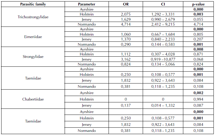
Table 2: Age as a risk factor associated with gastrointestinal infections caused by GIPs. Results are presented as OR and at a 95% CI.

Table 3: Variables as risk factors associated with gastrointestinal infections caused by GIP. Results are presented as OR and at a 95% CI.
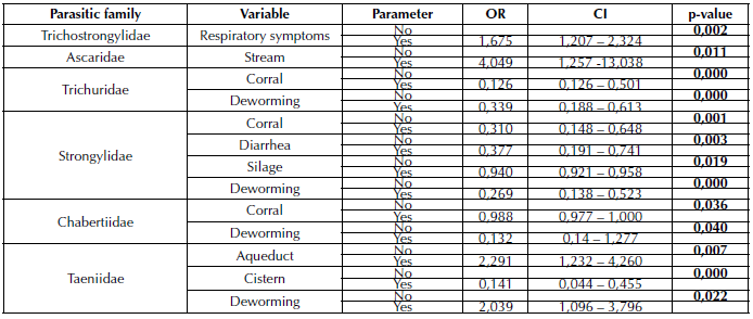
The analysis of variables such as sanitary management through deworming of the evaluated individuals showed a significant statistical association with the parasitic families Trichuridae, Strongylidae, Chabertiidae, and Taeniidae (p=0,000, p=0,000, p=0,040, and p=0,022, respectively), as well as a relationship between the implementation of the corral for cattle management and the presence of parasites of the Chabertiidae (p=0,036), Strongylidae (p=0,001), and Trichuridae families (p=0,000) (Table 3).
As for the water sources used in the productions, streams, aqueducts, and cisterns were found to have an association with Ascaridae and Taeniidae (p <0,05), whereas supplementation with silage was significant for Strongylidae (p <0,05).
Discussion
In Colombia, GIP prevalence values of 50,l5% (Pinilla-León et al., 2019) and 83,2% (Pinilla et al., 2018) have been reported, the latter being the closest to that reported in the central province of Boyacá. Likewise, Pinilla et al. (2019) determined a prevalence of 59% in Belén and 52,1% in Duitama (Boyacá), which is lower than the values obtained in this research. As for Latin America, in two districts of the Mantaro Valley (Perú), values of 24,5 and 30,3% have been reported (Briones-Montero et al., 2020), as well as a 39% prevalence in México (Fernández-Figueroa et al., 2015), in addition 47,8% for seasons with high rainfall and 46,2% for dry periods (Figueroa-Antonio et al., 2018). Prevalence values of 67,5% have also been reported in Perú (Colina et al., 2013), as well as 34,2% for Venezuela (Urdaneta-Fernández et al., 2011), with these being lower than those of this study.
Other regions of the world have reported prevalence values such as 51,5% in Pakistan (Shah et al., 2021), 60,46% in Iraq (Aram, 2020), 39,8% in Kombolcha city, Ethiopia (Ayele et al., 2020), 90,8% in Ghana (Squirere et al., 2019), 20,9% in Canada (Scott et al., 2019), 20,03% in India (Das et al. 2018), 40,6% in Germany (May et al., 2017), 38,8% in Indonesia (Nurtjahyani and Agustin, 2015), 86,9% in Taiwan (Huang et al., 2014), and 20,4 and 94,5% in Poland (Piekarska et al., 2013).
Among the possible factors that influence the variation of prevalence values are the high anthelmintic resistance developed by ruminants due to indiscriminate application of common antiparasitics (Geurden et al., 2014; Borges et al., 2015), climate factors that affect parasite biological cycles (Okulewicz, 2017), seasonal conditions that modify the intensity of parasite loads (Squire et al.,2019), and pre-seasonal rainfalls during the sampling phase that allow parasite development and dietary changes (Mahmood et al., 2014). Additionally, it should be considered that appropriate levels of humidity and temperature are required for the development of etiological agents and infective stages (Taylor et al., 2016), conditions that are more likely to occur in non-stabled farms (Squire et al., 2019).
The most prevalent families in cattle were Trichostrongylidae, Eimeriidae, and Taeniidae. However, in Venezuela, it was reported that the most frequent genera belong to the families Trichostrongylidae, Ancylostomatidae, and Strongylidae (Morales et al., 2012). Likewise, Sun et al. (2018) reported that the most prevalent parasites were Haemonchus, Ostertagia and Trichostrongylus, belonging to the Trichostrongylidae family, as well as Oesophagostomum of the Strongyloidae family. This variation may be due to environmental factors, the reproductive stage and sex of the animal, and the grazing and agricultural practices in farms. These factors play a determining role in the presence of infectious stages, thus favoring the development of reproductive cycles and the viability of eggs and larvae, which depend on the time of year, age, and immunological status of the host (Colina et al., 2013).
The age of the animals can also influence the parasite load, with the Strongyloididae family being the only one that showed statistical significance with this variable; individuals between 3 and 4 years old were less likely to get infected by parasites of the aforementioned family than bovines between 2 and 3 years old. This may be due to the fact that young cattle are susceptible to parasites, as demonstrated in previous research (Urdaneta-Fernández et al., 2011; Taylor et al., 2016; Briones-Montero et al., 2020). Therefore, keeping young animals in the same housing as older animals may expose them to parasitic infections (Pinilla et al., 2018; Squire et al., 2019). However, individuals in younger age groups may become more resistant to primary infection with some parasites as they reach maturity (Taylor et al., 2016).
Concerning the breed, contact with Holstein acts as a risk factor for infection by the Trichostrongylidae and Taeniidae families for the Ayrshire breed, which could mean that the former is more likely to suffer from parasitosis of these families than the Ayrshire breed cattle. However, the low amount of studies on parasites in dairy cows in tropical areas and their relationship with the breed of the animal hinders comparisons with the results obtained in this research. Da Silva et al. (2008) state that Holstein breed females, given their higher milk production, have a higher egg count per gram of feces than lower-production animals.
Since the response of ruminants depends on the nutritional status and metabolic requirements for milk production, there is a negative energy balance at the beginning of lactation. Therefore, there is a decrease in the expression of cytokines associated with the phenomenon of anthelmintic resistance (Pryce et al., 2000). Thus, specialized breeds (e.g., Holstein and Jersey) are more prone to suffer from GIPs because of their high production and their susceptibility to the stress of tropical climates, which decrease their immunological status, not to mention the management system used for the different breeds, which also causes stress (Silva et al., 2008).
The presence of in-farm pens acts as a protective factor against Trichuridae, Strongylidae, and Chabertiidae. Zootechnical factors play a significant role in the behavior of the incidence and intensity of parasitic invasion. The characteristics of the facilities and the area destined for the livestock farm, the type and form of feeding, the rearing system, and hygienic measures have a decisive influence on the conformation of the parasitological landscape of any herd (Soca et al., 2005).
Additionally, it should also be noted that deworming cattle is regarded as a protective factor against the presence of parasites of the Trichuridae, Strongylidae and Chabertiidae families, i.e., it is less likely that cattle will present these parasites, although, in the case of the Taeniidae family, deworming practices in production are considered to be a risk factor due to a condition that contradicts the biological relationship that may exist between the observed variables and the parasite family. Anthelminthic treatment is regarded as fundamental for helminth control in cattle given its ease of use, its relatively low cost, and the lack of effective alternative options (Woodgate et al., 2017). However, it should be taken into account that integrated parasite control entails harmony with helminths without them causing clinical conditions, as well as management strategies such as pasture rotation and selection of resistant and resilient animals, thus making it a more effective method to reduce parasitic infection levels (Bennema et al., 2010).
Regarding the water sources used for cattle, there is a negative relationship between streams and the Ascaridae family, probably as a risk factor, whereas, for the Taeniidae family, the presence of cisterns behaves as a protective factor, which does not agree with what was reported by Cornejo-Soto (2019), who found higher parasitic prevalence values in animals whose water consumption was supplied directly from ditches without receiving adequate potability treatment. Contaminated water is considered to be the main vehicle involved in the transmission of parasites. When ditches are used as a drinking water source, the possibility of GIPs in cattle increases, contrary to what occurs with an aqueduct system due to the physical-chemical processes performed to ensure optimal conditions for consumption (Cornejo-Soto, 2019).
Likewise, feeding practices contribute significantly to parasitosis, given that the oral route is the main access path for infective stages in the organism. Forages can constitute a source of infestation if they come from areas that have been fertilized with excreta or residues contaminated by infective stages of parasites (Soca et al., 2005; Henriques et al., 2021). This agrees with Urdaneta-Fernández et al. (2011), who state that the consumption of forage increases the risk of transmission of gastrointestinal nematodes, given that it is the main route of infection for most of the parasites that inhabit the intestinal tract. Thus, the consumption of silage acts as a protective factor against GIPs of the Strongylidae family since, in this case, feeding is not based solely on forage. In addition, the nutritional status of cattle can influence the severity of parasitic infection (Huang et al., 2014).
Conclusions
The results obtained in this research show the high presence of GIP in cattle of the central province of the department of Boyacá (95,6%), where the most prevalent families were Trichostrongylidae, Eimeriidae, Taeniidae, and Trichuridae. Likewise, age showed a significant statistical association with the Strongyloididae parasitic family. The breed variable of the individuals evaluated was associated with the Trichostrongylidae, Eimeriidae, Strongylidae, Chabertiidae, and Taeniidae families. The possible association of risk factors is related to the close relationship between the individuals and the parasitic families evaluated.
References
License
Copyright (c) 2022 Martin-Orlando Pulido-Medellin, Henry-Alexander Lopez-Buitrago, Diana-María Bulla-Castañeda, Diego-José García-Corredor, Adriana-María Díaz-Anaya, Julio-Cesar Giraldo-Forero, Rosa-Isabel Higuera-Piedrahita

This work is licensed under a Creative Commons Attribution-NonCommercial-ShareAlike 4.0 International License.
When submitting their article to the Scientific Journal, the author(s) certifies that their manuscript has not been, nor will it be, presented or published in any other scientific journal.
Within the editorial policies established for the Scientific Journal, costs are not established at any stage of the editorial process, the submission of articles, the editing, publication and subsequent downloading of the contents is free of charge, since the journal is a non-profit academic publication. profit.

